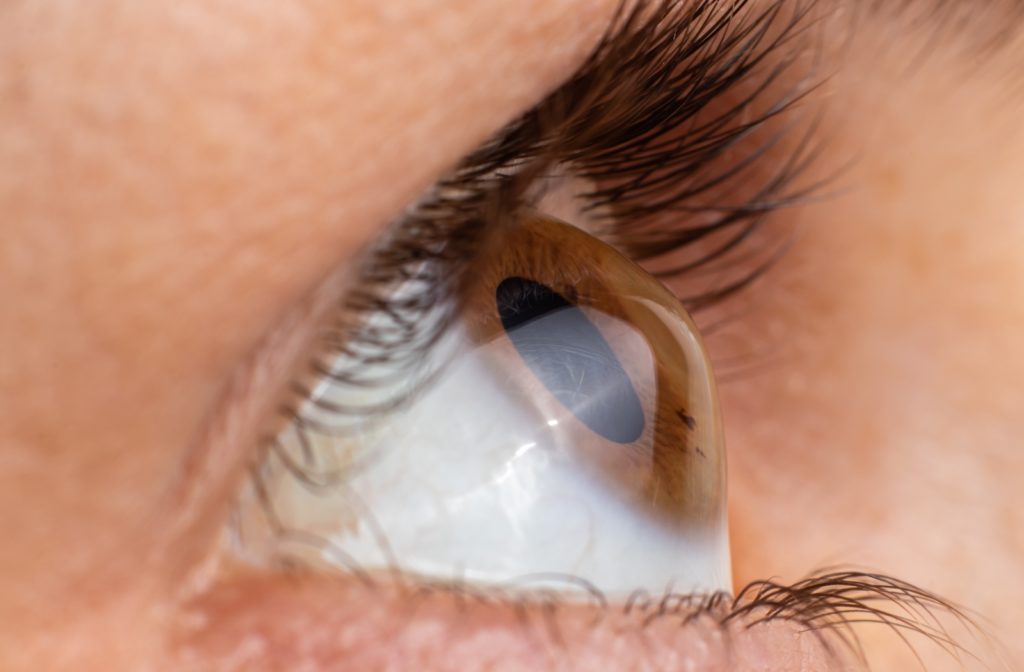
How does keratoconus develop?
The exact cause of keratoconus is still not known with certainty, but medical science has identified several risk factors. Genetic predisposition, biochemical disorders, environmental influences and even certain mechanical factors such as excessive rubbing of the eye may play a role.
During the course of the disease, the collagen fibres in the cornea weaken and are unable to maintain their regular shape, and the cornea gradually thins and bulges outwards. As a result, refraction becomes irregular, which can lead to serious vision problems.
Who is most often affected?
Keratoconus typically starts in late adolescence or early twenties and often affects both eyes – although not necessarily equally. The disease is more common:
- family accumulation,
- associated with allergic conditions (e.g. hay fever, atopic dermatitis),
- for severe eye rubbing,
- systemic diseases such as Down’s syndrome or Marfan syndrome.
It’s important to know that although keratoconus does not cause pain or inflammation, vision deterioration is progressively worse and can lead to permanent vision loss if left untreated.
What are the symptoms of keratoconus?
In the early stages of the disease, it is difficult to distinguish between other refractive errors, but certain symptoms may be particularly characteristic. It is worth paying attention to the following signs:
- gradually worsening vision that is no longer corrected by glasses,
- frequent dioptre changes (higher cylinder value) in spectacle controls,
- distorted vision: wavy, blurred outlines,
- photosensitivity,
- ghostly vision, especially at night,
- increased difficulty in seeing through contact lenses,
- sudden deterioration of vision that does not improve with rest or eye drops.
Diagnosis of keratoconus
A routine vision test is often not enough to detect keratoconus. To diagnose the disease, you need special tools and imaging tests, which our opticians have.
The most commonly used test methods:
- Corneal topography – detailed map of the cornea
- Pachymetry – measurement of the thickness of the cornea
- OCT (Optical Coherence Tomography) – Layer analysis and deep structure analysis
These modern techniques not only allow early detection, but are also excellent for monitoring the progress of the disease.
How can keratoconus be treated?
The treatment of keratoconus always depends on the progression of the disease and the patient’s visual acuity. Several options are available:
- Glasses or soft contact lenses – effective only in the early stages
- Contact lenses designed for a special shape – such as gas permeable or hybrid lenses
- Corneal cross-linking (CXL) – an advanced UV-light procedure that stabilises the cornea and can stop the disease from worsening
- Corneal implants (Intacs) – small semi-circular inserts that help to restore the cornea to a more regular shape
- Cornea transplant – last resort in severe cases
Living with keratoconus: what to look out for?
Although keratoconus can be challenging to diagnose, with proper treatment, vision can be stabilised in the long term. However, it is important to follow these guidelines:
- regular eye examinations,
- avoid rubbing the eyes,
- the correct use of prescribed equipment and treatments,
- wear sunglasses in bright light,
- proper management of allergic reactions, preventing eye irritation.

